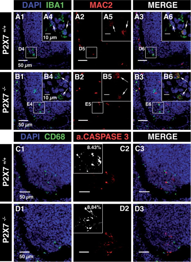
Figure 10.

Microglia activation and MN developmental cell death were not altered in the SC of E13.5 P2X7R−/− mouse embryos. A, B, Confocal image of immunostainings against IBA1 (A1 and B1) and Mac-2 (red; A2 and B2) in the SC of wild-type (A) and P2X7R−/− (B) mouse embryos. A1, B1, DAPI staining (blue) and microglia staining (IBA1 immunostaining, green) in the ventrolateral region of the SC. A3, B3, Superposition of IBA1 and Mac-2 immunostainings. Note that Mac-2 immunostaining colocalized with IBA1 immunostaining, indicating that microglia were activated in the LMC of the SC of E13.5 P2X7R+/+ mouse embryos (arrows; A4–A6) and of E13.5 P2X7R−/− mouse embryos (arrows; B4–B6). A4, B4, Single confocal section showing microglia located in the ventrolateral region of the SC. C, D, Confocal image of immunostainings against CD68 (C1 and D1) and activated caspase-3 (red; C2 and D2) in the ventral SC of wild-type (C) and of P2X7R−/− (D) mouse embryos. C1, D1, DAPI staining (blue) and microglia staining (CD68 immunostaining, green) in the ventrolateral region of the SC. C3, D3, Superposition of CD68 and activated caspase-3 immunostainings. Insets in C2 and D2 are measurements of the percentage of fluorescence in the ventrolateral region of the SC (see Materials and Methods).
