Figure 3.
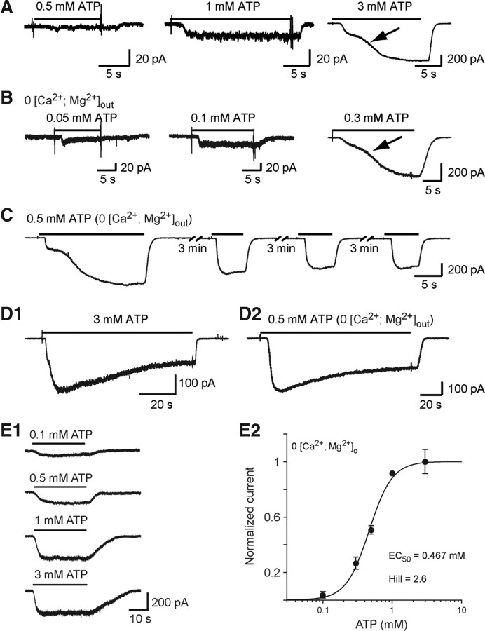
ATP evokes inward currents in embryonic microglia. A, Example of responses evoked by 0.5, 1, and 3 mm ATP in the presence of 1.3 mm [Ca2+]o and 3 mm [Mg2+]o. Note the biphasic activation phase (arrow) of the response evoked by 3 mm ATP. B, Currents evoked by 0.05, 0.1, and 0.3 mm ATP in [Ca2+ + Mg2+]o = 0. C, Currents evoked by repetitive applications of 0.5 mm ATP in [Ca2+ + Mg2+]o = 0. D, Current evoked by long application of 3 mm ATP in the presence (D1) and of 0.5 mm ATP in the absence (D2) of [Ca2+ + Mg2+]o. This response slowly desensitized (half-amplitude decay time = 26.9 s in the presence of [Ca2+ + Mg2+]o and 41.9 s in free [Ca2+ + Mg2+]o solution). Large inward currents evoked by 140 s application of ATP after eliciting a biphasic response slowly decreased with time in both normal [Ca2+, Mg2+]out (3 mm ATP) and free [Ca2+, Mg2+]out (0.5 mm ATP) solutions and reached a plateau representing 50.5 ± 3.1% (N = 5) of the peak current in normal [Ca2+, Mg2+]out solution and 64.7 ± 5.2% (N = 8) in free [Ca2+, Mg2+]out. The half-desensitization time was 25.9 ± 3.3 s (N = 5) in normal [Ca2+, Mg2+]out and 36.9 ± 6.5 s (N = 8) in free [Ca2+, Mg2+]out. E1, Example of currents evoked by increasing ATP concentrations after having obtained a biphasic response ([Ca2+ + Mg2+]o = 0). E2, Dose–response curve of ATP-evoked currents as shown in E1 (EC50 = 467 μm; Hill coefficient = 2.6). Each point represents the average of 5–10 measurements. The amplitude of the ATP-evoked responses has been normalized to the current amplitude of the responses evoked by 1 mm ATP (see Materials and Methods). (Vh =−60 mV). Error bars indicate SEM.

