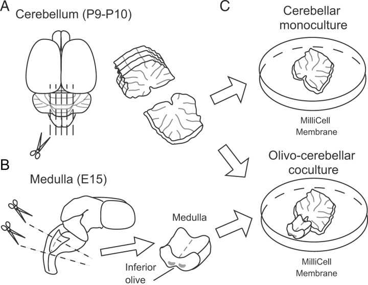
Figure 1.
Schematic diagram of the method for preparing cocultures of the cerebellum and the medulla. A, The cerebellum was dissected out from rats or mice at P9 or P10 under deep anesthesia. Cerebellar slices with 300 μm (for rats) or 250 μm (for mice) thickness were dissected from the vermis. Scissors represent the sites of sectioning. B, The whole brain was dissected after decapitation from each fetal rat at E15, and the medulla containing inferior olivary neurons was dissected. C, A block of the medulla was plated at the ventricular side of a cerebellar slice on a membrane filter that was coated with rat-tail collagen.

