
Figure 2.
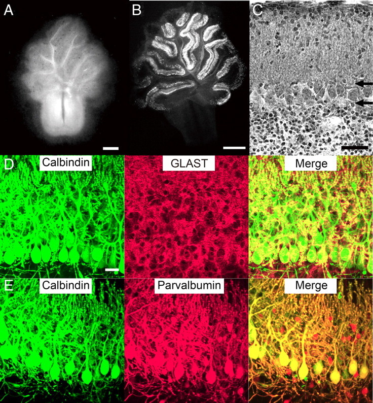
Cytoarchitecture in cocultures. A, Dark-field micrograph of a coculture at 3 weeks in vitro. B, Fluorescent immunohistochemistry using an antibody against the PC marker calbindin D-28K at 1 week in vitro. C, Nissl-stained section of the cultured cerebellar slice at 1 week in vitro. Horizontal arrows show the boundaries between the PC layer and the molecular layer (top arrow) or the granule cell layer (bottom arrow). D, E, Bergmann glia and inhibitory interneurons in cocultures. After 10 DIV, cocultures were double immunostained for calbindin (green) and GLAST, a marker of Bergmann glia (D; red), or for calbindin (green) and parvalbumin, a marker of inhibitory neurons (E; red). Note that GLAST signals are closely associated with calbindin signals and that calbindin-negative/parvalbumin-positive inhibitory neurons are present in the molecular layer and the PC layer. Scale bars: A, 0.5 mm; B, 1 mm; C, 50 μm; D (for D, E), 20 μm.