Figure 1.

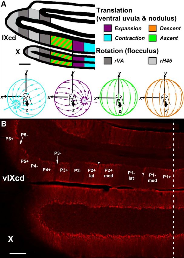
ZII organization and electrophysiological zones in the medial VbC (i.e., ventral uvula and nodulus) of pigeons. A shows the four functional classes of Purkinje cells responsive to patterns of optic flow resulting from self-translation in three-dimensional space: contraction (light blue), expansion (purple), ascent (green), and descent (orange) [based on the study by Wylie and Frost (1999)]. In the lateral VbC (i.e., flocculus), Purkinje cells are responsive to patterns of optic flow resulting from self-rotation about either the vertical axis (rVA) or an horizontal axes oriented 45° to the midline (rH45) as indicated by gray shading (Wylie and Frost, 1993; Wylie et al., 1993). B shows a coronal section through folia IXcd and X immunoreacted for ZII. The ZII stripes are numbered P1 to P7 (medial to lateral) from the midline (indicated by the dashed line). The ZII+/− pairs from P1+ to P5− are indicated in ventral IXcd as well as P6+. P6−, P7+, and P7− are found more rostrally (Pakan et al., 2007). P1− is divided into medial and lateral portions by a small satellite immunopositive band one to two Purkinje cells wide in the middle of P1− denoted “?.” P2+ is divided into medial and lateral portions by a small immunonegative “notch” in the middle of P2+ (see inverted triangle). Folium X does not have ZII stripes, as all Purkinje cells are ZII+. Scale bars: A, 500 μm; B, 300 μm.
