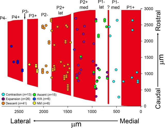
Figure 6.
Distributions of optic flow neurons in ZII stripes in the ventral uvula. This figure shows the recording sites of contraction (light blue), expansion (purple), ascent (green), descent (orange); cells not modulated to visual stimuli (yellow) and rVA (dark blue) cells from all cases are indicated. Both the caudorostral (y-axis) and mediolateral (x-axis) position are indicated. For the caudorostral axis, all measurements are relative to the most caudal section containing folium IXcd. For the medial–lateral axis, to permit comparisons between cases, the width of each ZII stripe was normalized (see text for more details). In total, 107 recording sites are indicated. The optic flow zones correspond to the ZII stripes as follows: contraction, P1+ and P1−med; expansion/ascent, P1−lat and P2+med; descent, P2+lat and P2+. The six cells that were not modulated (NM) by visual stimuli were all localized to P3+. Consistent with Pakan et al. (2011), the six rVA cells were localized to P4+.

