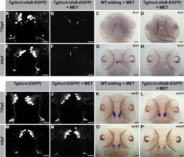
Figure 1.
Establishment of inducible HCRT neuron-ablated transgenic larvae. Confocal fluorescence (left) and stereomicroscopic ISH (right) imaging of representative larvae (dorsal view with head pointing to the top). As indicated by confocal imaging of EGFP expression, MET treatment induced HCRT cell-ablation in 72 hpf and 6 dpf Tg(hcrt:nfsB-EGFP) larvae (B, F). Hcrt (C, G, D, H) and mch (K, L, O, P) mRNA ISH pattern in 72 hpf and 6 dpf larvae, all with MET treatment. MET treatment caused specific cell ablation in HCRT neurons (D, H) and had no effect on adjacent MCH neurons (L, P). HCRT-neuron ablation was not induced in the absence of nfsB (C, G, J, N) or MET (A, E). In A, arrows and arrowheads indicate the soma of HCRT neurons and projecting axons, respectively. In B, D, F, and H, arrows indicate cell residues after ablation. Scale bars: A, B, E, F, I, J, M, N, 50 μm; C, D, G, H, K, L, O, P, 250 μm.

