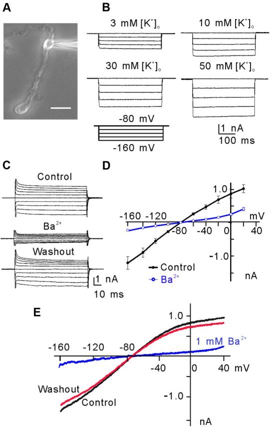
Figure 1.

Inwardly rectifying K+-selective current (Kir) in rat retinal Müller cells. A, Micrograph showing a typical isolated rat Müller cell. Scale bar, 10 μm. B, [K+]o dependence of hyperpolarization-activated Kir currents of Müller cells. The currents were evoked by a series of hyperpolarized voltage pulses from a holding potential of −80 mV in increments of −20 mV. Note that the current amplitudes were increased with an increase in [K+]o. C, Ba2+ (1 mm) blocked the membrane currents of a Müller cell, voltage clamped at −80 mV and stepped to −160 mV in −10 mV increments and then to +20 mV in 10 mV increments. D, Membrane currents plotted against the step potentials. Note the K+-selective weakly rectifying Kir4.1-like I–V relationship (n = 10). The Kir currents were reduced significantly by Ba2+ (1 mm). E, Currents elicited by a 1 s voltage ramp from −160 mV to +40 mV before (control), after the application of Ba2+ (1 mm) and washout of Ba2+.
