Figure 2.

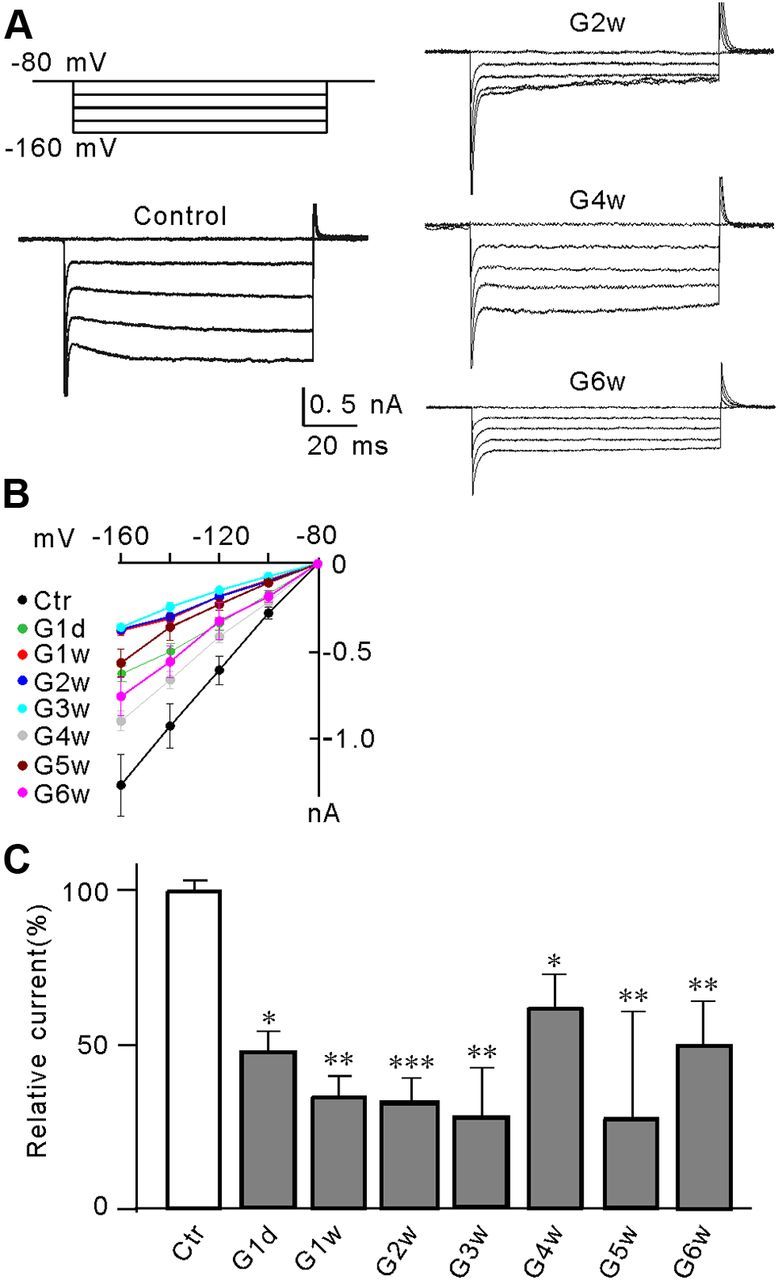
Suppression of Kir currents in Müller cells in a rat COH model. A, Voltage-clamp analysis of the Kir current changes at negative membrane potentials. Cells were clamped at −80 mV and stepped to −160 mV in −20 mV increments (top left). Whole-cell membrane currents were recorded in Müller cells isolated from sham-operated control (Ctr) and COH rats on the second (G2w), fourth (G4w), and sixth week (G6w) after the operation. B, The I–V relationships, showing voltage-dependent suppression of Kir current amplitudes in Müller cells obtained from Ctr and COH rats at the first day after surgery (G1d), and the first to sixth week (G1w–G6w) after the operation C, Summarized data showing that the average Kir current peak amplitudes decreased when the IOP was elevated. All data are normalized to control. Error bars represent SEM. n = 8∼15, *p < 0.05, **p < 0.01, and ***p < 0.001 versus control (Ctr).
