Figure 6.
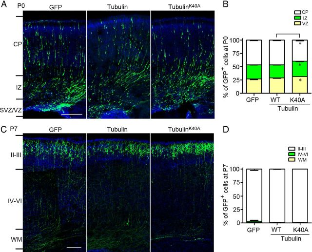
TubulinK40A overexpression causes a slight migratory defect in the cortical projection neurons. A, B, Coronal sections of the cerebral cortex at P0 (A) and P7 (C) in rats electroporated at E16.5 with a GFP-expressing plasmid along with tubulin or tubulinK40A. The sections were counterstained with DAPI (blue). The histograms show the distribution of transfected cortical neurons in electroporated brain at P0 (B) and P7 (D) across different cortical regions. Scale bars: 200 μm. *p < 0.05 versus tubulin in the corresponding region. All data represent the means ± SEM.

