Figure 7.

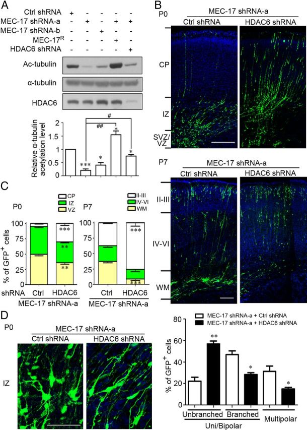
Loss of HDAC6 reduces the defects in MEC-17-deficient cortical projection neurons. A, Immunoblots and histogram showing the acetylation level of α-tubulin in ND7–23 cells transfected with different constructs as indicated. *p < 0.05, ***p < 0.001 versus control shRNA; #p < 0.05, ##p < 0.01 versus MEC-17 shRNA-a. B, C, Coronal sections of rat cortex at P0 (B, top) and P7 (bottom) electroporated at E16.5 with a GFP-expressing plasmid along with MEC-17 shRNA-a or MEC-17 shRNA-a together with HDAC6 shRNA. Sections were counterstained with DAPI (blue). Histograms show the distribution of transfected cortical projection neurons in electroporated brain across different cortical regions at P0 (C, left) and P7 (right). **p < 0.01, ***p < 0.001 versus those coexpressing MEC-17 shRNA-a with control shRNA in the corresponding region. D, Coronal sections of rat cortex in the IZ region and histogram showing the distribution of neurons expressing MEC-17 shRNA-a or MEC-17 shRNA-a and HDAC6 shRNA at the multipolar stage, unipolar/bipolar stage (unbranched), or unipolar/bipolar stage with highly branched leading processes (branched) at P0. *p < 0.05, **p < 0.01 versus those coexpressing MEC-17 shRNA-a with control shRNA. Scale bars: B, 200 μm; D, 50 μm. All data represent the means ± SEM.
