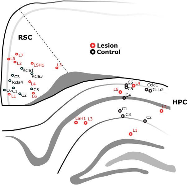
Figure 3.
Schematic diagram showing the representative anteroposterior position and anatomical locations of all recording electrodes in the study (30 electrodes in 18 animals). LFP recordings were made from the septal CA1 subregion of the HPC and the septal RSC. Animal identification labels are provided to permit identification of the proportion of the animals that recorded from both regions simultaneously (n = 11) and those that were recorded from either region singularly (n = 7).

