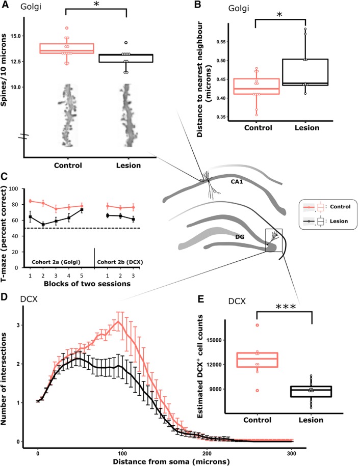
Figure 7.
MTT lesions were associated with a decrease in both number (A) and clustering (B) of spines in the basal dendrites of Golgi-stained CA1 pyramidal neurons in Cohort 2a. Performance on a T-maze task was impaired in MTT lesion animals relative to controls in Cohort 2a and Cohort 2b (C). DCX is a marker of adult neurogenesis; in Cohort 2b, MTT lesions resulted in a decrease in the complexity of DCX+ neurons (D) as well as a decrease in the stereologically estimated overall number of DCX+ neurons in the dentate gyrus (DG; E). Cohort 2a (lesion, n = 9; controls, n = 12); Cohort 2b (lesion, n = 12; control, n = 8). Central schematic diagram represents the morphology and anatomical location of neurons that were sampled. *p < 0.05; ***p < 0.001.

