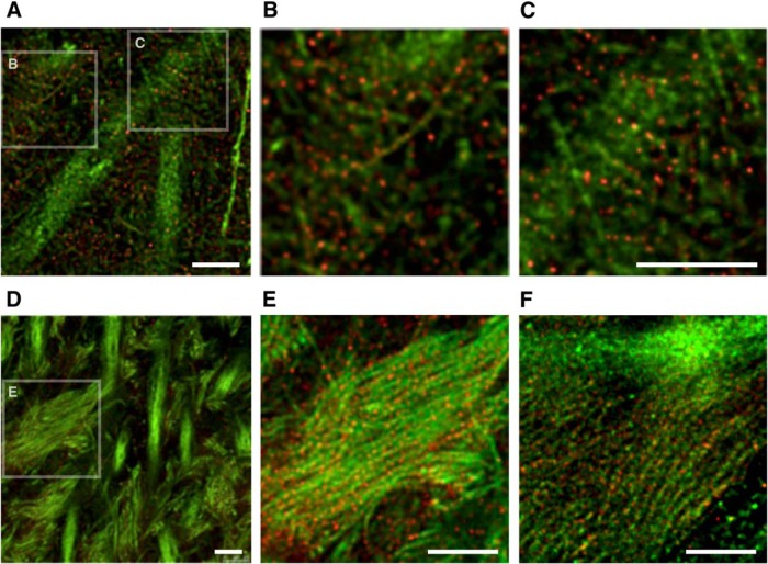
Figure 6.
High-resolution imaging of tau in the hippocampal region of non-Tg mouse brains. A–C, Hippocampal CA1 in non-Tg mice labeled with DM1A (green) and anti-tauN (red) and imaged using STED. The boxes in A are the areas that are magnified in B and C. In CA1, tau is discontinuously localized in axons. D, Hippocampal CA3 in non-Tg mice labeled with DM1A (green) and anti-tauN (red). The box in the left image is the area that is magnified in E. Punctate tau labeling was observed on mossy fiber axons but was not found in the apical dendrites of pyramidal neurons in area CA3. E, Magnified image of CA3 labeled with DM1A (green) and tauN (red). F, Magnified image of CA3 labeled with DM1A (green) and RTM38 (red). Scale bars, 2.5 μm.

