Figure 7.
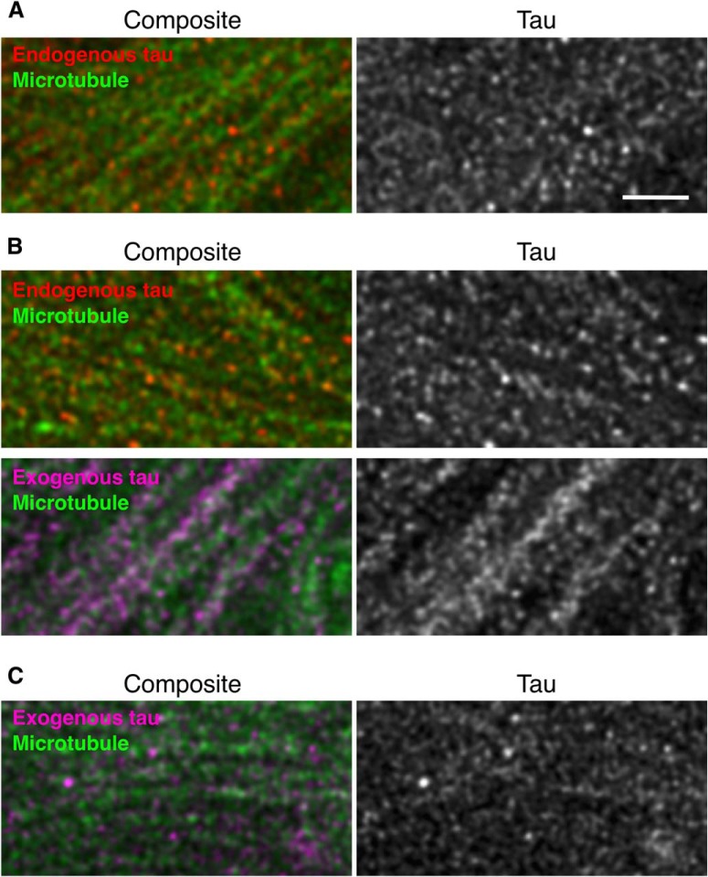
High-resolution imaging of endogenous and exogenous tau in the hippocampi of Tg and KI mouse brains, Mossy fibers of non-Tg (A), P301L-Tg (B), and heterozygous V337M-KI (C) mice were labeled with DM1A (green) and anti-RtauN (endogenous tau, red) or RTM49 (human tau, magenta) and observed using STED microscopy. Merged views (left) and tau signal (right) were shown. Exogenous tau in Tg mice, but not in KI mice, showed a more continuous localization pattern. Scale bar, 1 μm.

