Figure 9.
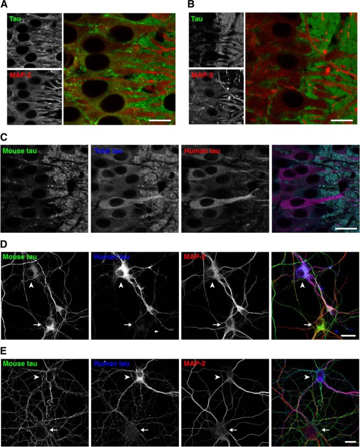
Localization of tau in P301L-Tg mice during development. A, Brain sections from P7 non-Tg mice were subjected to immunostaining for tau (anti-tauN, green) and MAP2 (HM2, red). There were some overlapping signals of tau and MAP-2 in the somata. Scale bar, 20 μm. B, Brain sections from P14 non-Tg mice were immunostained for tau (anti-tauN, green) and MAP2 (HM2, red), which exhibited interdigitated patterns. Scale bar, 20 μm. C, Brain sections from P14 P301L-Tg mice were subjected to immunostaining for mouse tau (anti-RtauN, green), total tau (tauN, blue), and human tau (Tau12, red). Scale bar, 20 μm. D, E, Mixed primary cultures of hippocampal neurons from the brains of both non-Tg and P301L-Tg mice were immunostained with anti-mouse tau (RTM47, green), anti-human tau (tau12, blue), and anti-MAP2N (red) at 3 DIV (D) and 14 DIV (E). The arrowheads indicate cells that were derived from P301L-Tg mice and the arrows indicate cells from non-Tg mice. Scale bars, 20 μm.

