Figure 2.
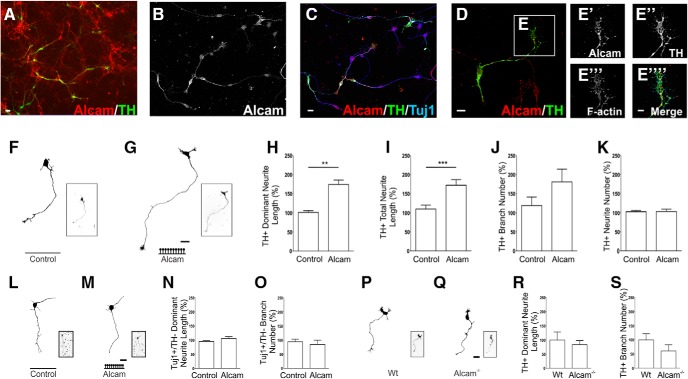
Alcam in vitro expression and function during dopaminergic axonal growth. Representative images of neurons in primary midbrain cultures immunolabeled for Alcam (A–C) show Alcam expression in dopaminergic and nondopaminergic neurons. Nondopaminergic neuronal populations display a range of expression from very strong, to completely absent. In dopaminergic neurons, punctate Alcam expression was present in cell bodies and axons (D) and in F-actin+ growth cones (E). Representative images and corresponding axonal traces of TH+ neurons in primary midbrain cultures grown on control (F), or high-density Alcam substrate (G). Quantification of axon growth in the presence of an Alcam substrate resulted in a significant increase in length of the longest (dominant) neurite (H) (unpaired t test, t = 4.533, df = 8, p = 0.0019, n = 5) and the length of all (total) neurites per neuron (I) (unpaired t test, t = 2.659, df = 8, p = 0.0289, n = 5). No significant effect was observed on number of branches (J) or number of neurites (K) per neuron. Representative traces of nondopaminergic midbrain neuron traces (Tuj1+/TH−) from primary midbrain neurons cultured on control (L) or high-density Alcam substrate (M). Quantification of axon growth in the presence of Alcam substrate resulted in no significant effect on neurite length (N) or branch number (O) (n = 5). Representative axonal traces of dopaminergic (TH+) neurons in primary midbrain cultures from WT (P) and Alcam−/− mice (Q). Quantification of axon growth showed no significant changes due to loss of Alcam function on (R) neurite length or (S) branch number (unpaired t test, n = 4). Scale bars: A–D, 20 μm; E, 10 μm; F, G, L, M, P, Q, 20 μm. Data are shown as mean ± SEM. **p < 0.01, ***p < 0.001.

