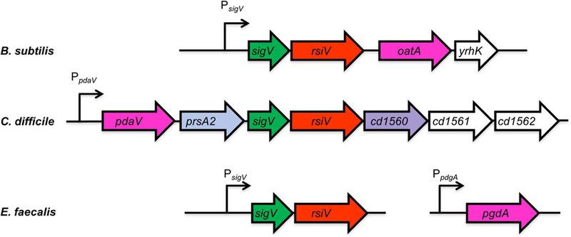
Figure 1. Alignment of sigV regions from B. subtilis, C. difficile and E. faecalis.

Shown in green is sigV (σV), rsiV (anti-σ factor RsiV) is red, the genes encoding peptidoglycan modifying enzymes are in pink (pdaV, pgdA and oatA), prsA2 (peptidylprolyl isomerase) is light blue and an rsiV ortholog cd1560 which lacks the σ factor binding domain is purple, the genes in white have no known function. The C. difficile genes are numbered based on C. difficile 630 numbering.
