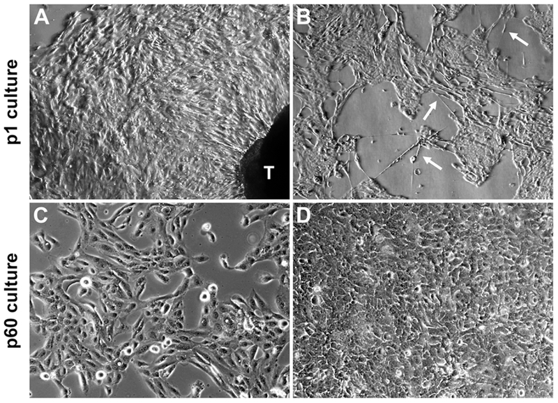
Figure 1. Epithelial cells from ex vivo tissue culture of clinical prostate tumor specimens.
Representative results illustrate the common features from ex vivo culture of three matched pairs of patient specimens. A, in primary culture (p1) outgrow of cells with epithelial morphology from prostate tissue dice (labeled as T) became obvious after 7 days of ex vivo culture (40×). B, the p1 epithelial cell culture contained at least two different cell types, cobblestone-like cells interspersed with long neuroendocrine-like cells (arrowheads, 40×). C, a few HPE-15 cells from primary culture survived continuous passaging as a cell line. Low density HPE-15 cells at passage 60 (p60) (100×). D, high density HPE-15 culture at p60 (100×).

