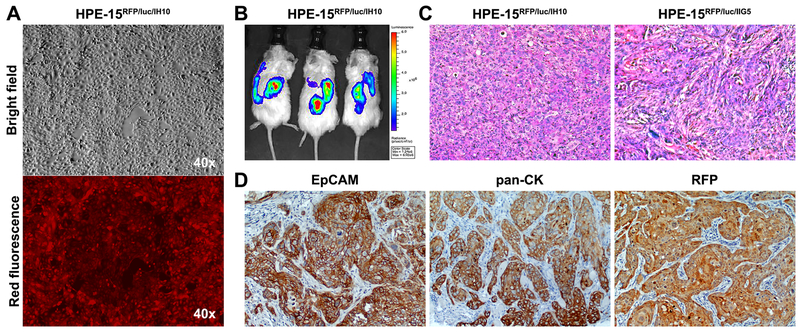
Figure 3. Acquisition of tumorigenicity through interaction with highly tumorigenic PCa cells.
HPE-15RFP/luc cells, recovered from 3-D co-culture with two aggressive PCa cells, IH10 or IIG5, were found to have acquired tumorigenic potential by s.c xenograft tumor formation. A, photograph of HPE-15RFP/luc cells isolated from 3-D co-culture with IH10 cells and amplified under puromycin selection conditions (HPE-15RFP/luc/IH10). No contaminating IH10 cells were seen since all the cells were red fluorescent. B, representative BLI results of NSG mice bearing s.c HPE-15RFP/luc/IH10 tumors. C, representative H&E staining of s.c xenograft tumors of HPE-15RFP/luc/IH10 and HPE-15RFP/luc/IIG5 cells isolated from 3-D co-culture with metastatic IH10 or IIG5 cells in mice (200×). D, representative IHC images of HPE-15RFP/luc/IH10 tumors, which expressed pan-CK and EpCAM epithelial markers. Strong RFP staining confirmed the origin of the tumor formation to be from HPE-15RFP/luc/IH10 cells (100×).

