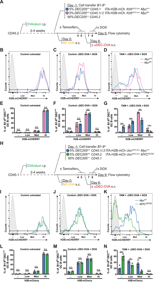
Figure 6. MYC is both necessary and sufficient for GC B cell division.
(A) Schematic representation of the experimental protocol for (B-G). (B-D) Representative histograms show H2B-mCherry fluorescence among B1–8hiR26ERT2-CreMyc+/+tTA–H2B– mCh (blue) and B1–8hR26ERT2-CreMycfl/+tTA–H2B–mCh (pink) GC B cells in mice treated as indicated. (E-G) Quantification of (B-D). Control mice in (B) and (E) are untreated with TAM, αDEC-OVA and DOX. Summary of results from 3–6 mice in two independent experiments. Mean percentage of H2B-mCherry negative, low, medium or high B1–8hiR26ERT2-CreMyc+/+tTA–H2B–mCh and B1–8hiR26ERT2-CreMycfl/+tTA–H2B–mCh GC B cells. (H) Schematic representation of the experimental protocol for (I-N). (I-K) Representative histograms show H2B-mCherry fluorescence among B1–8hiUbcERT2-CreMyc+/+tTA–H2B–mCh (blue) and B1–8hiUbcERT2-CreR26StopFLMYC tTA–H2B–mCh (MycOE/OE, green) GC B cells in mice treated as indicated. (L-N) Quantification of (I-K). Control mice in (I) and (L) are untreated with TAM, αDEC-OVA and DOX. Summary of results from 3–5 mice in two independent experiments. Mean percentage of H2B-mCherry negative, low, medium or high B1–8hiUbcERT2-CreMyc+/+tTA–H2B–mCh and B1–8hiUbcERT2-CreR26StopFLMYC tTA–H2B–mCh GC B cells. Solid grey represents non-fluorescent cells. DOX = Doxycyclin hyclate. Unpaired two-tailed student’s t test. N.S.: p > 0.05 (not statistically significant); *p < 0.05, **p < 0.01, ***p < 0.001.

