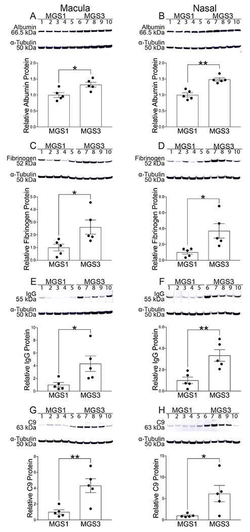
Fig. 2:

Western analysis of post mortem NSR protein from macula with corresponding pixel density graphs (A, C, E, G) and from nasal periphery with corresponding pixel density graphs (B, D, F, H) obtained from the Minnesota Lions Eye Bank. Immunoblots were done with the following antibodies: albumin (A,B), fibrinogen (C,D), IgG (E,F), C9(G,H). Loading control α-tubulin (50kDa) bands are shown below each set of lanes. Graphs show band densitometry normalized to loading control calculated using Image J software. Numbers represent mean values (±SEM). MGS1 (n=5) indicates normal, MGS3 (n=5) indicates intermediate AMD. Statistical analysis was performed using student’s two-tailed unpaired t-test. p<0.05, ** p<0.01.
