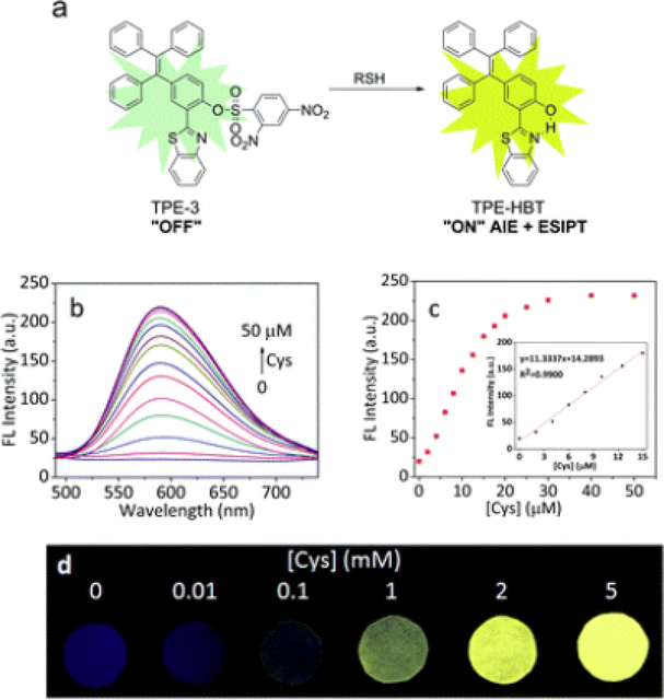
Fig. (6).
(a) Proposed sensing mechanism for TPE-3 and biothiols. (b) Fluorescence spectra of TPE-3 (10 μM) upon addition of Cys (0–50 μM) in PBS buffer (10 mM, pH = 7.4, containing 45% DMSO). (c) Fluorescence response of TPE-3 at 589 nm to Cys concentration (0 – 50 μM). Inset: linear range for Cys detection. Spectra were recorded after incubation with different concentrations of Cys for 15 min, λex = 370 nm. (d) Fluorescent photographs of TPE-3 deposited on test papers after immersed into buffer solutions (10 mM, pH = 7.4) with different concentrations of Cys under a UV lamp (365 nm) (reproduced with permission from Chen et al. [83] Copyright © The Royal Society of Chemistry, 2017).

