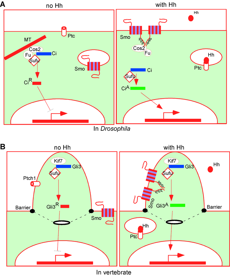
Figure 1.
The outlines of Hh signaling
(A) In Drosophila, Ptc keeps Smo inactive, partly by promoting its internalization and degradation. Full-length Ci is sequestered in the cytoplasm in a complex with Cos2, Fu and Sufu, whereas Ci repressor (CiR) inhibits target gene expression in the nucleus. Hh blocks Ptc function, allowing the phosphorylation and dimerization/oligomerization of Smo, leading to Ci activation (CiA) and target gene expression. MT: microtubule;
(B) In vertebrates, Ptch1 in the primary cilia prevents Smo ciliary localization and activation. Gli3 enters the cilia with Sufu and Kif7 and is processed into Gli3R. Shh binds Ptch1, leading to Smo ciliary translocation, phosphorylation, dimerization/oligomerization and Gli3 activation (Gli3A). A membrane barrier near the base of the cilia prevents free diffusion of Smo.

