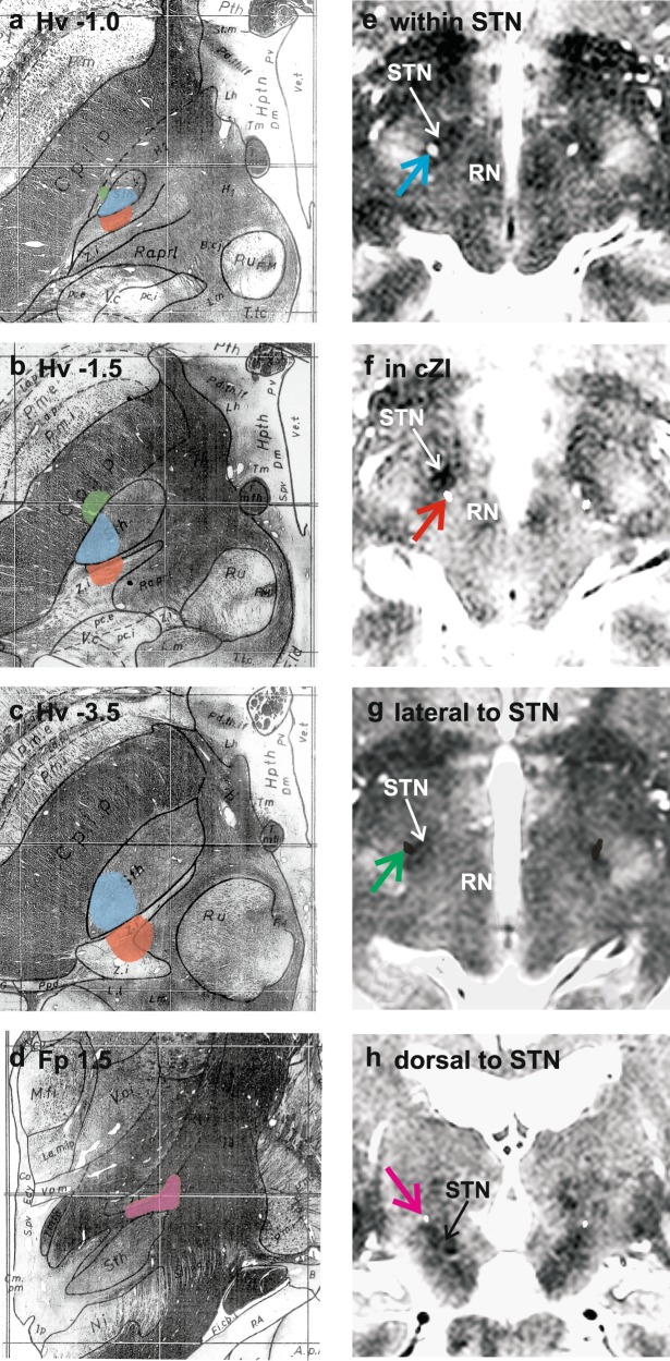
Fig. 1.
Anatomical locations of the active contacts were divided into four groups, the site and extent of each indicated by coloured shading in representative labelled example sections (a–c axial, d coronal) from the stereotactic atlas of Schaltenbrand and Wahren21: within STN (blue); dorsal to STN (purple); posteromedial to STN, in cZI (red); and lateral to STN (green). e–h Typical examples of images used to localise electrodes is presented for each active contact location. The intra-operative study is fused with the planning MRI scan in the plane of the active contact centroid. The stylette artefacts (coloured arrows; colours as in a–d) from the intra-operative imaging (CT study in e, f and h; MRI study in g) are thus visible on the anatomical planning scans and allow contact localisation. e–g show axial and h coronal slices. STN and red nucleus (RN) are labelled

