Fig. 4.
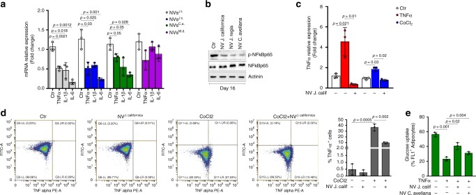
NVs reduce TNF-α levels and increase glucose uptake in adipocytes. a Cytokines mRNA expression was analyzed through RT-qPCR in hypertrophic adipocytes treated with NVs isolated from Juglans regia (J.r.), Juglans californica (J.c.), Corylus avellana (C.a.), and Malus domestica (M.d). b p-NFkBp65 protein levels were analyzed in hypertrophic adipocytes treated with NVs isolated from Juglans regia, Juglans californica, and Corylus avellana. Immunoblots reported are representative of three independent experiments. Actinin was used as a loading control. Uncropped images are shown in Supplementary Fig. 7. c, d TNF-α mRNA expression (c) and intracellular TNF-α protein (d) levels were analyzed by RT-qPCR and cytofluorimetry, respectively, in adipocytes treated with TNF-α or CoCl2 in combination with NVs isolated from Juglans californica. The gating strategy of cytofluorimetric analysis is shown in Supplementary Fig. 8. e Glucose uptake was measured by flow cytofluorimetry in insulin-stimulated hypertrophic adipocytes treated with NVs isolated from Juglans californica and Corylus avellana. Data are expressed as means ± SD (n = 3)

