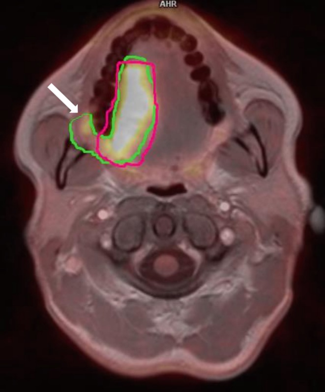
Fig. 2.

Primary tumor volume (GTV, gross tumor volume) delineated with manual method and presented on fusion of 18-fluorine-labeled fluorodeoxyglucose positron emission tomography (PET) and magnetic resonance (MRI) images. PET-based GTV (green line) are larger than MRI-based GTV (pink line) and include tumor’s infiltration on the retromandibular triangle (arrow)
