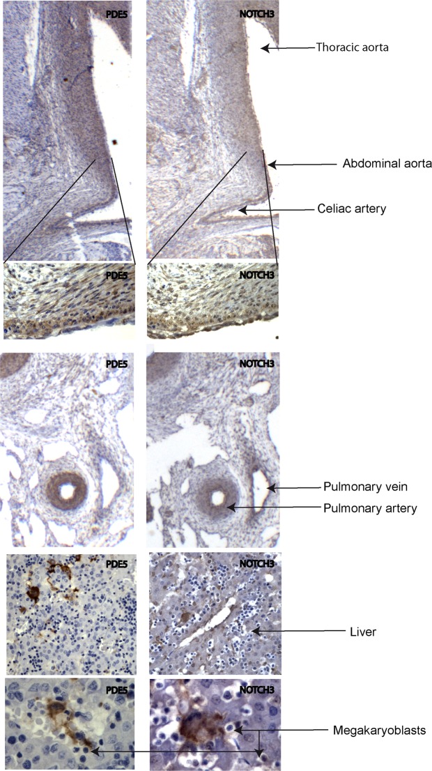
Figure 5.
PDE5 and NOTCH3 expression during development of mouse and human fetal aortas. Representative immunohistochemical evaluation of PDE5 and NOTCH3 on early mouse (12 days post coitum, dpc) and human (7 weeks post-conception, wpc) embryos showing positivity for both protein in the medial layer of the dorsal aorta, pulmonary arteries, gastrointestinal tract, and in megakaryoblasts of the liver in both mouse and human embryos. Black arrows indicate thoracic aorta, abdominal aorta, celiac artery, pulmonary vein and pulmonary artery respectively.

