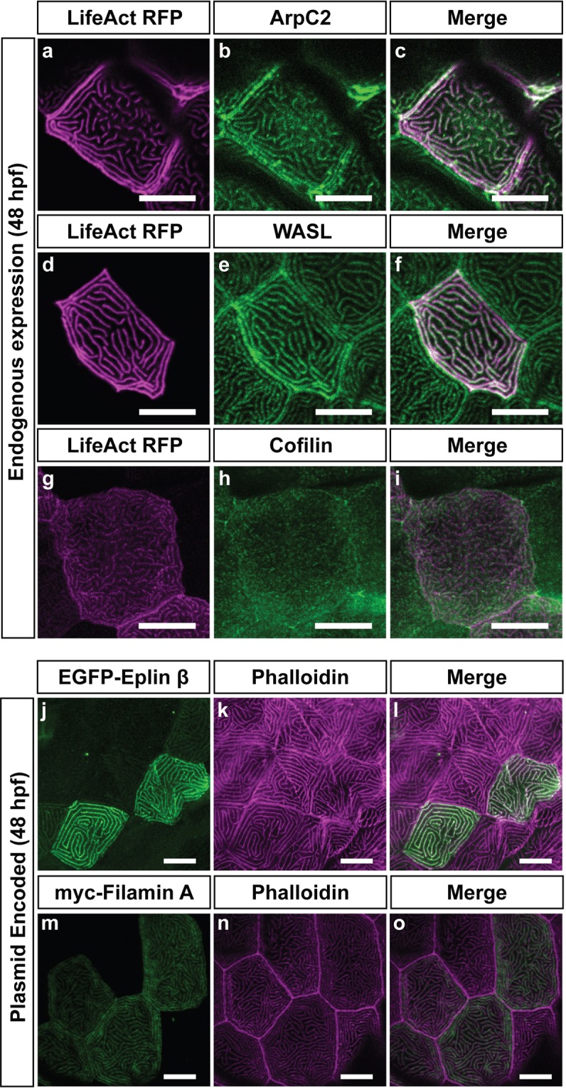
Figure 3.

Localization of actin binding proteins at the microridge at 48 hpf. Immunolocalisation of ArpC2 (b), WASL (e), and Cofilin (h) at 48 hpf with LifeAct-RFP in magenta that marks F-actin (a,d,g) at 48 hpf. Respective merges are shown in (c,f,i). Plasmid encoded Eplin-β (j), and Filamin A (m) in green localize to microridges marked with phalloidin (k,n) in magenta in respective merges (l,o). Plasmids were injected at the 1 cell stage and expressed in a clonal fashion. While all other images are obtained from the head epidermis, Eplin images are taken on the flank. Scale bars are 10 µm.
