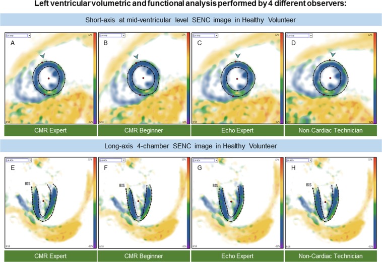
Figure 2.
Example of fast-SENC images acquired in a healthy volunteer and uploaded into a dedicated MyoStrain software. CMR images were derived in three long-axis and three short-axis views. Endocardial and epicardial borders were traced at end-diastolic and end-systolic cardiac phases by four observers: CMR expert (A,E) CMR beginner (B,F) echocardiography expert (C,G) and non-cardiac technician (D,H). CMR = cardiac magnetic resonance; SENC = strain-encoded imaging.

