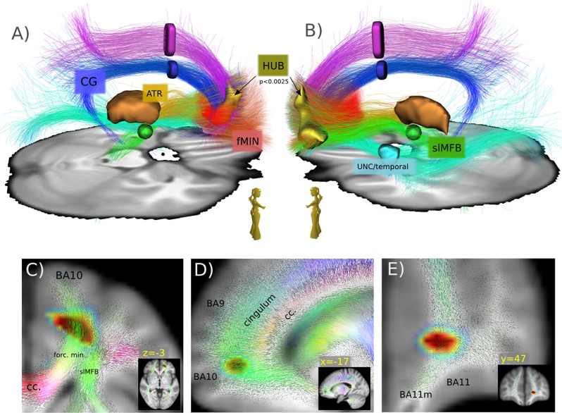
Fig. 4. Qualitative depiction of the involved bundles.
A qualitative view a, b of the major bundles involved in the HUB region. Tractography from a HCP group template, seeded from HUB (p < 0.01). Tracts are further separated (ROIs). Anterior thalamic radiation (ATR), superolateral medial forebrain bundle (slMFB), forceps minor (FMIN), cingulum (CG), uncinate fascicle (UNC), inferior fronto-occipital fascicle (IFOF), and superior anterior fascicle (SAF). c–e colored quiver plots give a prototypical impression of the local white matter geometry in the neighborhood of the HUB region

