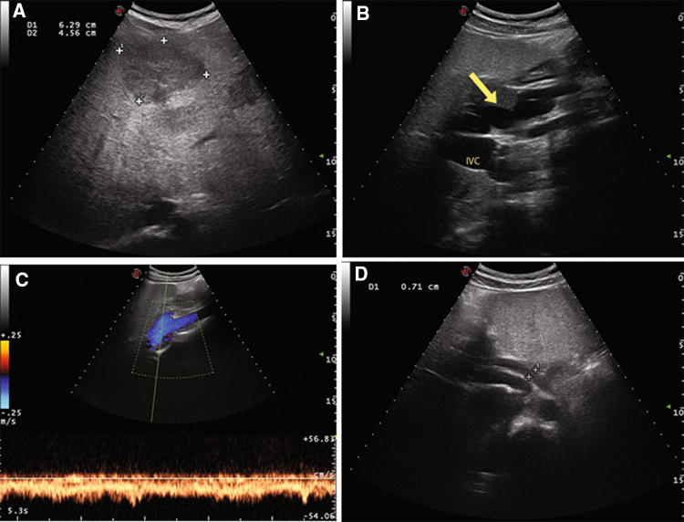
Fig. 1.
Case 2 patient. Color-Doppler Ultrasound of the liver showing multiple patchy hypoechoic liver nodules, the main one of 6.3 cm of diameter in the III–IV segment (a), a tortuous splenomesenteric trunk (arrow) with a turbulent flow apparently directed towards inferior vena cava (IVC) in the absence of main intrahepatic portal branches (b, c), hepatic artery hypertrophy (1D)

