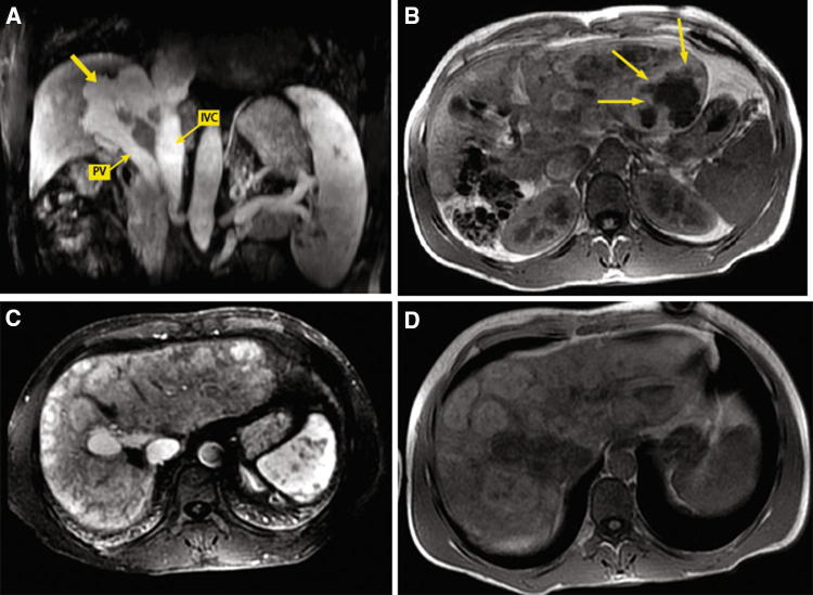
Fig. 4.
Case 3 patient. T1-Weighted fat-saturated magnetic resonance images obtained during the portal venous phase showing a tortuous vascular shunt (arrow) between the main portal trunk (PV) and the inferior vena cava (IVC) in the absence of intrahepatic PV branches (a); an expansive multinodular mass of the left lobe with areas of internal colliquation (arrows) (b) and multiple nodules up to 5 cm in the right lobe with contrast enhancement persistent in late phase (c). T1-weighted fat-saturated hepatobiliary phase magnetic resonance image showing that hepatic lesions are hyperintense or have a hyperintense peripheral rim (d)

