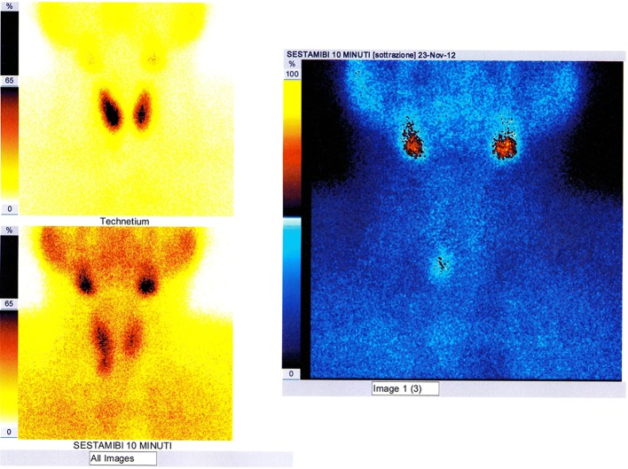
Fig. 10.
Image of planar scintigraphic examination performed with either “single-tracer double-phase” technique or with “double tracer subtraction” technique, documenting uptake by a right inferior parathyroid adenoma, which had already been visualized by the CDHR-NUS (see Fig. 3)

