Fig. 5.

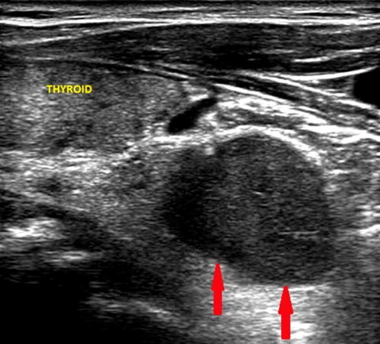
Ultrasound image of a left inferior parathyroid adenoma (indicated by the red arrows), located near the pole of the thyroid lobe with partial retrosternal localization and of the maximum (longitudinal) diameter of about 2.3 cm

Ultrasound image of a left inferior parathyroid adenoma (indicated by the red arrows), located near the pole of the thyroid lobe with partial retrosternal localization and of the maximum (longitudinal) diameter of about 2.3 cm