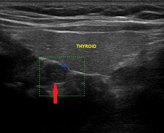
Fig. 8.

Ultrasound image of retrothyroidal exophytic thyroid node (indicated by the red arrow) with a maximum diameter of about 7 mm, which simulates a superior pathological parathyroid; the color-Doppler examination shows poor peripheral vascularization
