Figure 5.

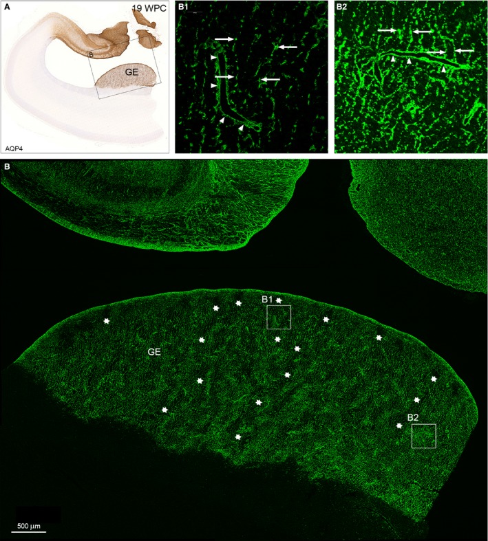
Distribution of AQP4 in the ganglionic eminence from a 19 wpc human fetal brain. A neighboring section to that of hippocampus and adjacent temporal cortex in Fig. 4K immunostained for AQP4 for bright‐field light microscopy (A) and confocal laser scanning microscopy (B). The framed area in (A) includes the ganglionic eminence (GE) in the lateral temporal wall and part of the strongly reacting hippocampal formation facing the lateral ventricle in the medial wall, which will be dealt with in Fig. 6. The GE in (B) is characterized by an uneven distribution of AQP4‐positive radial glial fibers resulting in a sponge‐like compartmentalized structure characterized by an intricate system resembling tunnels with thin and thick walls. The interior of some of the worm‐like tortuous tunnels is indicated by rows of white asterisks and shown in higher magnification in (B1). The more dense structure of the walls is depicted in (B2). (B1) Regularly spaced thin radial glial fibers with evenly spaced varicosities (arrows) characterize the tortuous tunnels of the AQP4‐positive ganglionic eminence. Side branches of the radial glial fibers ensheathe blood vessels (arrowheads) as astroglial end feet, although they are still part of the radial glial fiber system. (B2) The matrix of the sponge‐like structure possesses a more dense network of thick, rather coarse radial glial fibers with larger and more intensively stained varicosities (arrows), many of which seem to terminate on blood vessels as astroglial end feet (arrowheads). GE, ganglionic eminence. Scale bar: (B) 500 μm.
