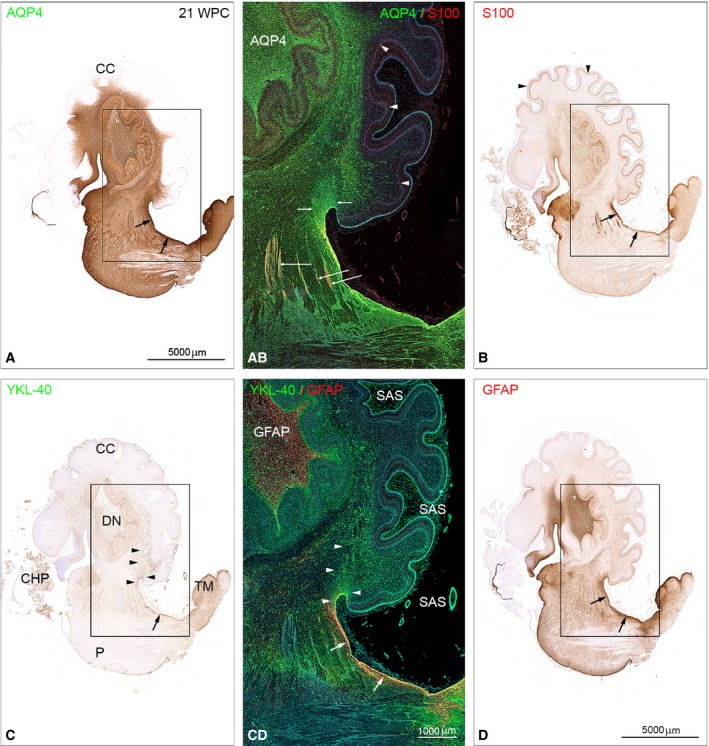
Figure 8.

Distribution of AQP4, S100 and YKL‐40, GFAP in brainstem and cerebellum from a 21 wpc (CRL: 200 mm) human fetal brain. Adjacent sagittal sections immunostained with antibodies against AQP4, S100 (A,B and AB) and YKL‐40, GFAP (C,D and CD). Note the marked differences in staining patterns with the entire brainstem and the core of cerebellum showing strong AQP4 immunoreactivity in contrast to a lack of staining of the cerebellar cortex (CC) in (A), whereas Bergman glia in cortex (arrowheads) in (B) exhibits a pronounced staining for S100. (C) A stream of YKL‐40‐positive cells migrates from the gliogenic hook between the dorsal brainstem and rostral cerebellum towards the center of cerebellum (arrowheads). All four astroglial markers show an overlapping pattern of immunoreactivity in the dorsal subpial gliogenic region (small black arrows) extending from the tectum mesencephali (TM in C) towards the cerebello‐mesencephalic junction. The boxed areas in (A‐D) are shown in (AB) and (CD) as double‐immunofluorescent sections stained with the same four antibodies and with nuclei labeled with DAPI shown in blue. The double‐labeled sections were subjected to whole‐slide fluorescent scanning and representative panels are displayed. (AB) and (CD) clearly demonstrate that GFAP‐, S100‐, AQP4‐ and YKL40‐positive cells represent separate although partly overlapping cell populations in the dorsal subpial gliogenic region (white arrows in CD). The radial glial cells of cerebellum (Bergmann glia) are strongly positively stained for S100 (arrowheads in AB and B) but not for the other astroglial markers. Cell aggregations seen along fiber tracts connecting cerebellum and upper brainstem seem to be distinctly immunoreactive for S100 (long white arrows in AB). The fiber tracts are surrounded by AQP4‐positive small cells (green). The migrating cell population from the dorsal subpial gliogenic region that is positively stained for YKL‐40 in (CD) (marked with white arrowheads) seems to be immunoreactive also for AQP4 (small white arrows in AB). Note the strongly GFAP‐immunoreactive core of the dentate nucleus (DN) in (CD) and a similar reactivity for AQP4 in AB. In the subarachnoid space (SAS) the numerous small leptomeningeal cells (green) and the smooth muscle cells (green) in small arteries are also YKL‐40‐positive. CC, cerebellar cortex; CHP, choroid plexus of the 4th ventricle; DN, dentate nucleus; P, pons; SAS, subarachnoid space; TM, tectum mesencephali. (A‐D) Same magnification. Scale bars: (A) 5000 μm; (AB,CD) same magnification; (CD) 1000 μm.
