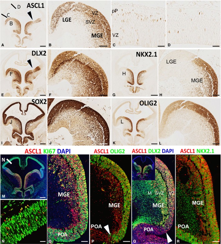
Figure 2.

Expression of ASCL1, DLX2, and associated proteins at 6.5 PCW. (A) Shows high expression of ASCL1 in ganglionic eminences, preoptic area, and hypothalamus. Location of origin of panels (B,C,D) are marked. Arrowhead indicates boundary between LGE and MGE. (B) Shows ASCL1 expression was high in the ventricular zone (VZ) of both LGE and MGE and also high in the subventricular zone (SVZ) of the MGE. In the dorsal telencephalon expression was low, but higher laterally (C) than dorsally (D) with a few cells deep in the VZ but more present at the boundary of the VZ and the nascent post‐mitotic preplate (pP). (E) Shows high expression of DLX2 in ganglionic eminences, preoptic area, and hypothalamus with lower expression in the VZ of the ganglionic eminences and higher expression in the SVZ and overlying mantle. NKX2.1 expression was confined to MGE, preoptic area and parts of the hypothalamus (G,H). SOX2 expression marked all progenitor cells in the forebrain and illustrates the extent of the SVZ in the MGE (I,J) and strong expression of OLIG2 also delineated the MGE (K,L). Immunofluorescence double‐labelling showed that ASCL1+ cells of the telencephalon expressed cell division marker KI67 and thus were dividing neuroprogenitors (M–O). Co‐expression patterns of ASCL1 with OLIG2 (P) and DLX2 (Q) define the boundary between MGE and preoptic area (POA), although NKX2.1 is expressed in both regions (R). Scale bars: 500 μm (A,E,I,G,K,M), 100 μm (B,F,H,J,L,O,P,R for Q see P or R), 50 μm (C,D,N).
