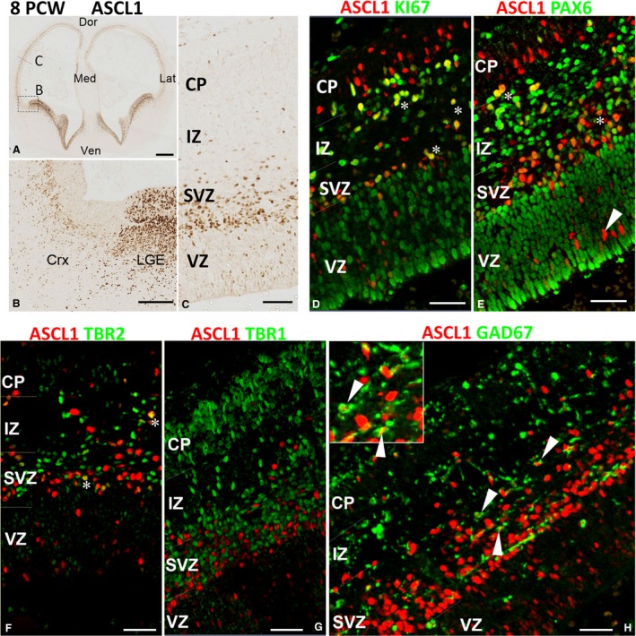
Figure 3.

ASCL1 expression in the cortex at 8 PCW. (A) Anterior section of telencephalon; box shows the approximate location of panel (B) and the line of panel (C). Very high expression of ASCL1 in the ganglionic eminences and septum, and discernible expression in the cerebral cortex was observed (A,B). In the cortex, ASCL1 was predominantly expressed in subventricular zone (SVZ) and intermediate zone (IZ) with (D) many cells co‐expressing the marker for cell division KI67 (yellow, e.g. asterisks) and (E) the marker for radial glia, PAX6 (yellow, asterisks) in these locations. However, in the ventricular zone (VZ), co‐expression with either marker was rare (arrowhead). (F) ASCL1 was also co‐expressed with the marker of intermediate progenitor cells TBR2 (asterisks) but not with TBR1 (G) the marker for post‐mitotic neurons. (H) Co‐expression with GAD67, marker of GABAergic interneurons (arrowheads). Inset shows two examples of these double‐labelled cells at higher magnification. Dor, dorsal; Med, medial; Ven; ventral; Lat, lateral; CP; cortical plate. Scale bars: 500 μm (A), 100 μm (B), 50 μm (C–G) and 30 μm (H).
