
Figure 4.
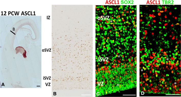
ASCL1 expression in the cortex at 12 PCW. (A) Section of telencephalon including MGE and LGE (high ASCL expression) with area B showing approximate location of panel (B). (B) Shows low expression of ASCL1 in the VZ but higher in the inner subventricular zone (iSVZ) and also throughout the outer subventricular zone (oSVZ). (C) Asterisks mark examples of double‐labelling of ASCL1 (red) with SOX2 (green, expressed by radial glia) and (D) mark examples of double‐labelling with TBR2 (expressed by intermediate progenitor cells). Scale bars: 1 mm (A), 100 μm (B) and 50 μm (C,D).