Figure 7.

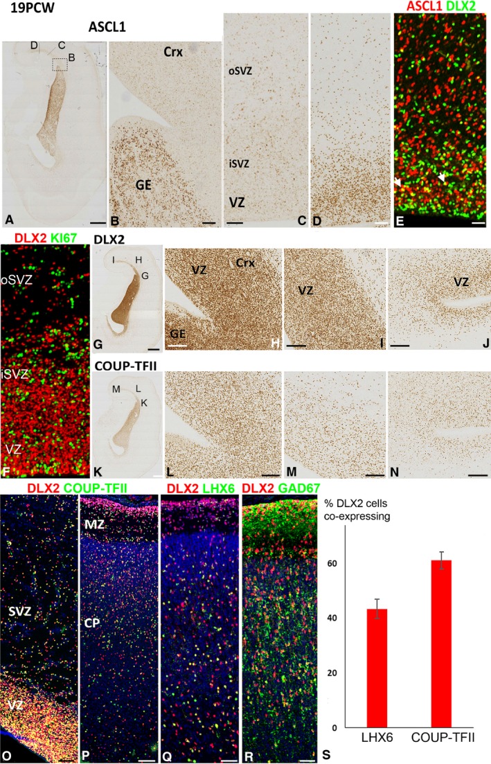
ASCL1 and DLX2 expression at 19 PCW. (A) Coronal section of cortex and ganglionic eminence immunostained for ASCL1 showing approximate location of panels (B,C,D). (B) High expression in the ganglionic eminence (GE) and very low expression in the lateral cortex which increases at more dorsomedial cortical locations (C,D) in both the inner and outer subventricular zone (iSVZ, oSVZ), but especially in the ventricular zone (VZ). (E) Co‐expression of ASCL1 (red) and DLX2 (green) was rare but observed in a few cells (yellow, e.g. arrowheads). (F) However, no co‐expression of DLX2 (red) with KI67 (green) was observed in the cortex. DLX2 expression was highest in the GE and in the lateral cortex close to the pallial/subpallial border, particularly in the VZ (G,H). More dorsomedially, the concentration of DLX2+ cells decreased (I,J). COUP‐TFII exhibited a similar pattern of expression (K–N), and DLX2 (red) and COUP‐TFII (green) were widely co‐expressed (yellow) throughout the cortical wall (O,P). A proportion of DLX2 cells also co‐expressed LHX6 (yellow, Q) and extensively co‐expressed GAD67 (R). At this stage of development, a higher proportion of DLX2+ cells co‐expressed COUP‐TFII than LHX6 (S). Scale bars: 2 mm (A,G,K), 200 μm (B), 25 μm (C–E for F see E) 200 μm (L,M,N) and 50 μm (O–R).
