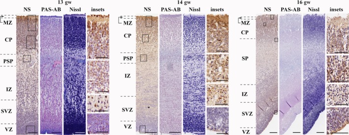
Figure 2.

Neuroserpin expression in the early second trimester (frontal lobe). Neuroserpin‐ip cells were present through the cortical plate and subplate. They had long processes and elongated perikarya showing migratory morphology. Minimal expression was seen in the marginal zone. Neuroserpin‐ip cells were observed in the ventricular zone of the lateral and caudal ganglionic eminences. MZ, marginal zone; CP, cortical plate; PSP, pre‐subplate; SP, subplate; IZ, intermediate zone; SVZ, subventricular zone; VZ, ventricular zone. Scale bars: (13th gw) NS, PAS‐AB, Nissl: 100 μm, insets: 50 μm; (14th–16th gw) NS, PAS‐AB, Nissl: 200 μm, insets: 50 μm.
