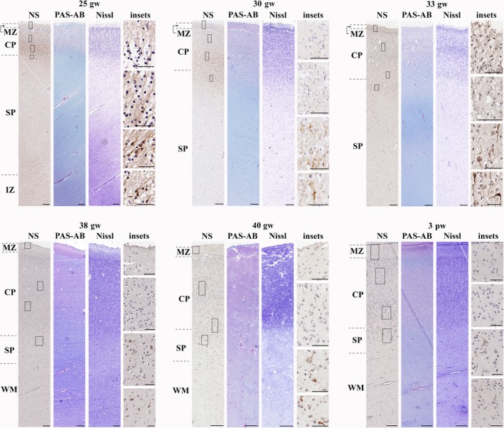
Figure 4.

Neuroserpin expression from the late second trimester until the first postnatal month (frontal lobe). Neuroserpin‐ip cells were situated to the deep cortical plate and subplate with a pyramidal and elongated shape. The upper and middle regions of the cortical plate were devoid of neuroserpin immunoreactivity. MZ, marginal zone; CP, cortical plate; SP, subplate; IZ, intermediate zone; WM, white matter. Scale bars: (25th gw–3rd pw) NS, PAS‐AB, Nissl: 200 μm, insets: 50 μm.
