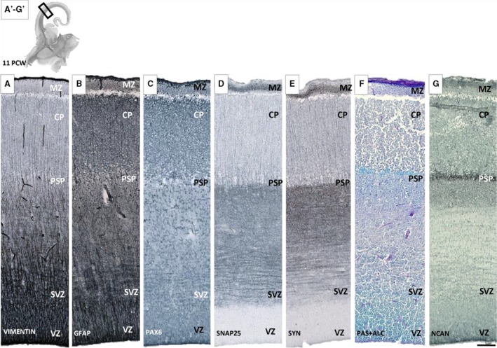
Figure 2.

Presubplate (PSP) monolayer shown with different glial (A,B,C), synaptic (D,E) and extracellular matrix (F,G) markers on sequent coronal sections of brain aged 11 postconceptional weeks (PCW). The most obvious delineation of PSP is visible on PAS+Alcian (F) and NCAN section (G), while on sections reacted for synaptic markers such as SNAP‐25 (D) and synaptophysin (E), immunoreactivity for synaptic markers is somewhat stronger in the presubplate layer and gradually merges with moderate (‘fibrillar‐like’) reactivity of deeper layers [intermediate (IZ) and subventricular zone (SVZ)]. Glial markers vimentin (A) and GFAP (B) show a modest change in geometry at the PSP level. Rectangles on the low magnification image (A′‐G′) correspond to the position of enlarged sequential sections below. CP, cortical plate; MZ, marginal zone; SVZ, subventricular zone; VZ, ventricular zone. Scale bar: 100 μm.
