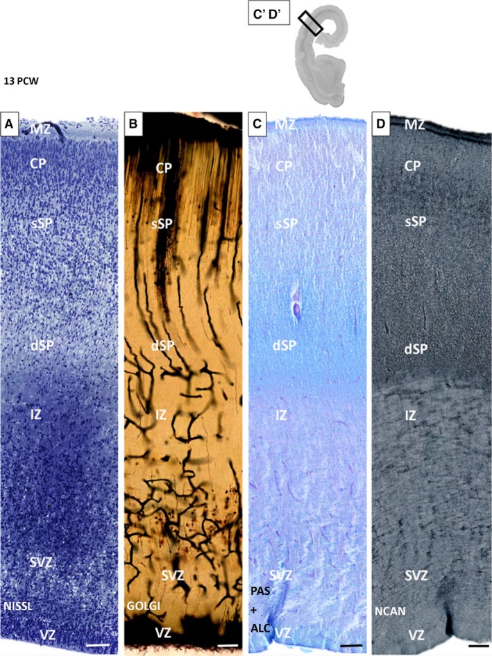
Figure 3.

Subplate (SP) formation phase. Bilaminar expanded SP shown on histological and immunocytochemical preparations on an old human neocortex at 13 postconceptional weeks (PCW). Loss of the deep portion of the cortical plate (CP; superficial ‘upper’ SP‐sSP) and gradual merging with the presubplate (deep ‘lower’ SP; dSP) are characteristic of the bilaminar SP. Below the SP is an intermediate zone (IZ) with darker staining due to the osmification of fibers on a 1‐μm plastic section prepared for electron microscopy (A). Prominent changes in orientation of radial glia fibers and vessels within the dSP are visible on Stensaas Golgi‐impregnated sections (B). Distribution of extracellular matrix markers PAS (C) and NCAN (D) shows a gradual increasing tendency towards the deep (lower) SP (D). MZ, marginal zone; SVZ, subventricular zone; VZ, ventricular zone. Rectangles on (C′‐D′) correspond to the position of enlarged sequential sections (C) and (D). Scale bar: 100 μm.
