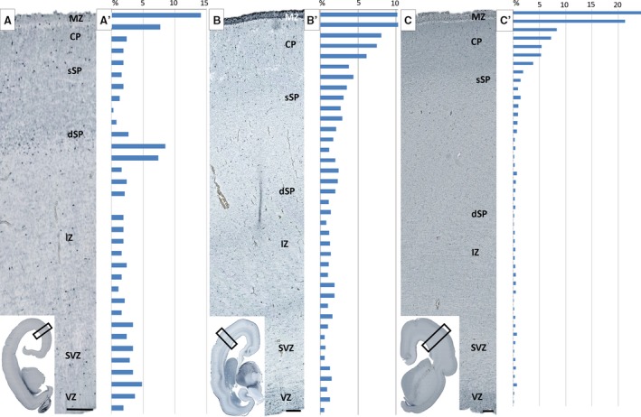
Figure 4.

Histograms show distribution of calretinin‐reactive neurons in 13 (A,A′), 15 (B,B′) and 17 (C,C′) postconceptional weeks (PCW) old specimens. The height of each bin in the histograms represents 50 μm of ventricle–pia distance. Histograms are juxtaposed to histological sections on the left side with indicated laminar compartments. Note higher concentration of calretinin cells in the deep lamina of SP (dSP) during SP formation (A′) and increased number of calretinin cells in the cortical plate (CP) and marginal zone (MZ) during midgestation. Cells are counted in observed surface areas of 1.3 mm2 in 13 PCW, 2.85 mm2 in 15 PCW, and 5.45 mm2 in 17 PCW specimens, and number of cells is expressed as a percentage noted above (see Materials and methods). IZ, intermediate zone; SVZ, subventricular zone; VZ, ventricular zone. Scale bar: 100 μm.
