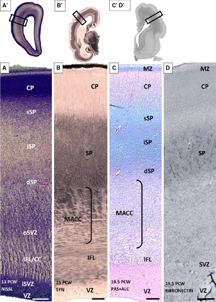
Figure 5.

Trilaminar subplate (SP) organization shown on Nissl (A) synaptophysin (B), PAS‐Alcian (C) and fibronectin (D) immunoreacted coronal sections of 13 (A′), 15 (B,B′) and 19.5 (C,C′,D,D′) postconceptional (PCW) brains. Three SP laminas [superficial (sSP), intermediate (iSP) and deep (dSP)] have no sharp borders but are recognizable due to different cell packing density (A) on the Nissl‐stained section. The most obvious sublamination is evident on PAS‐Alcian stained sections (C). (C′D′) Section used as an example for the approximate position (rectangle) of sequent (C,D) sections. (A′,B′,C′,D′) Rectangles mark the positions of the enlarged images (A,B,C,D). CC, corpus callosum; CP, cortical plate; dSP, deep SP; IFL, inner fibrillar layer; ; iSP, intermediate SP; MACC, multilaminar axonal‐cell layer; MZ, marginal zone; oSVZ, outer SVZ; SP, superficial SP; SVZ, subventricular zone; VZ, ventricular zones. Scale bar: 200 μm.
