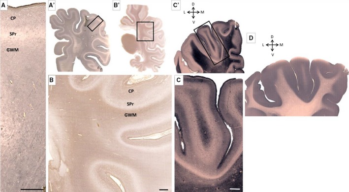
Figure 12.

Characteristic distribution of extracellular matrix marker chondroitin sulfate (A–C) and synaptophysin (D) at the newborn age. Subplate remnant (SPr), seen as wavy unstained line, along the hemisphere (A,A′,B,B′,C,C′) is in contrast to moderately stained underlying gyral white matter (GWM) and cortical plate (CP). Changes in synaptophysin distribution are very prominent and immunoreactivity involves both SPr and CP (D). Rectangles in (A′, B′ and C′) mark approximate positions on (A, B and C), respectively. Scale bar: 1 mm.
