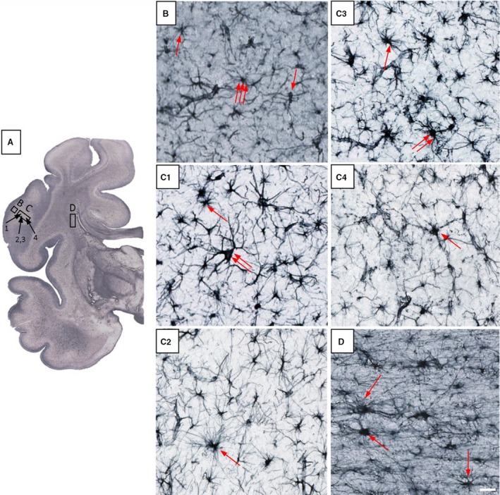
Figure 13.

Glia architectonics in 38 postconceptional weeks (PCW) old brain shows clear differences between cortical plate (CP; B), subplate remnant (SPr; C1–C4) and fetal white matter – capsula interna (D). SPr shows typical astrocytes (C1,C2 – one arrow), some giant neuron‐like GFAP‐reactive cells (C1 – two arrows) and prospective protoplasmic astrocytes (C3 – two arrows; C3,C4 – single arrow). Identification of fibrous astrocytes is possible due to their radiating unbranched processes, whereas protoplasmic astrocytes are not easy to identify because they show immature, partially branched processes. Rectangles marked with (B, C and D) indicate approximate positions of cells shown on higher magnified images, juxtaposed to the right. Scale bar: 25 μm.
